
Latissimus Dorsi
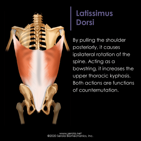
The latissimus dorsi produces adduction, extension, and medial rotation of the humerus and ipsilateral rotation of the thorax (counternutation). Acting like a bowstring, it may increase the thoracic kyphosis (counternutation).
Proximal Attachment: Lesser tubercle of the humerus and intertubercular groove
Distal Attachment: Spinous processes from T6 to L3, inferior scapular angle and, through the thoracolumbar fascia and lateral raphe of the thoracolumbar fascia, to the iliac crest and sacrum [1]. Down to L3, the fibers are attached to the spinous processes. Below L3, the fibers, as part of the superficial lamina of the thoracolumbar fascia, cross to the opposite side to attach to the sacrum, posterior iliac spine, and median sacral crest, and mesh with the fibers coming from the contralateral gluteus maximus [2].
Functions:
- Adduction, extension, and especially medial rotation of the humerus [3]p534 [1]p811
- In lifting, it acts to pull the object closer to the body.
- Pulls the shoulder posteriorly and causes ipsilateral rotation of the thorax [4]p518 [5]p327
- Because it crosses from the posterior to the anterior aspect of the trunk, it may cause flexion of the upper thoracic spine, acting like a bowstring, increasing the thoracic kyphosis (counternutation) [4]p172
- Thoracic extension (counternutation) from T5 – L3
- Compresses the sacroiliac joint contralaterally, enhancing stiffness [2, 6]
During gait, right trunk rotation increases the left lumbar lordosis [7]. Since gait causes reciprocal movements bilaterally, as the left lordosis increases (nutation), the right lumbar lordosis would decrease (counternutation).
By flattening the lumbar lordosis and increasing the thoracic kyphosis, it demonstrates that the lumbar and thoracic curves move in opposite A/P directions; when one increases, the other decreases. Because the lumbar and cervical curves move in the same A/P directions, it can be seen that, as the cervical and lumbar lordosis increase, the thoracic kyphosis decreases, and vice versus.
References:
- Standring, S., et al., eds. Gray’s Anatomy. 40th ed. 2008, Churchill Livingstone: London.
- Vleeming, A., et al., The posterior layer of the thoracolumbar fascia. Its function in load transfer from spine to legs. Spine, 1995. 20(7): p. 753-8.
- Warwick, R. and P.L. Williams, eds. Gray’s Anatomy. 35 ed., ed. R. Warwick and P.L. Williams. 1973, W.B. Saunders Company: New York.
- Oatis, C.A., Kinesiology. The Mechanics and Pathomechanics of Human Movement. 2004: Lippincott Williams & Wilkins.
- Neumann, D., Kinesiology of the Musculoskeletal System. Foundations for Physical Medicine. 2002: Mosby.
- Wingerden, J.v., et al. Muscular Contribution to Force Closure: Sacroiliac Joint Stabilization In Vivo. in 4th Interdisciplinary World Congress on Low Back & Pelvic Pain. 2001.
- Gracovetsky, S. Locomotion – Linking the Spinal Engine with the Legs. in Proceedings of the Second Interdisciplinary World Congress on Low Back Pain. 1995. San Diego, CA.